Swiss premium oral care

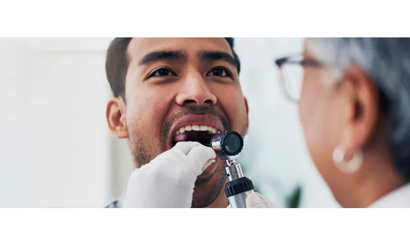
Why are teeth X-rayed at the dentist?
The reason for X-rays at the dentist becomes immediately apparent when we look at the anatomy of human teeth: Only about one third of the tooth is visible. The remaining two thirds are embedded in the gums and jawbone and cannot be examined with the naked eye. X-ray technology is used so that dentists can nevertheless detect abnormalities - both in the treatment of diseased teeth and as a preventive measure. Prophylactic x-rays can detect inflammation in the tooth and other changes before pain occurs.
X-rays of the teeth are used in the following areas:
- Recognising inflammation in living and dead teeth (e.g. inflammation of the tooth root)
- Diagnosis of periodontitis and other changes in the jawbone
- Detection of caries (also in spaces, under crowns and fillings)
- Detection of cracks and fractures
- Detection of cysts and tumours
- Planning of dentures
- Measuring the tooth length and checking the filling for root canal treatments
- Planning of oral surgery procedures
- Checking the condition of the wisdom teeth
What do you see on the x-ray?
During an X-ray, the teeth are illuminated for a short time with electromagnetic waves, known as X-rays. These rays then hit an X-ray film in the case of analogue X-rays and an image plate or sensor in the case of digital X-rays and image the teeth in full length. As dense tissue such as bones and teeth only allow a few rays to pass through, they appear light-coloured on the X-ray image. The darker an image area is, the more radiolucent it is - air, for example, is black.
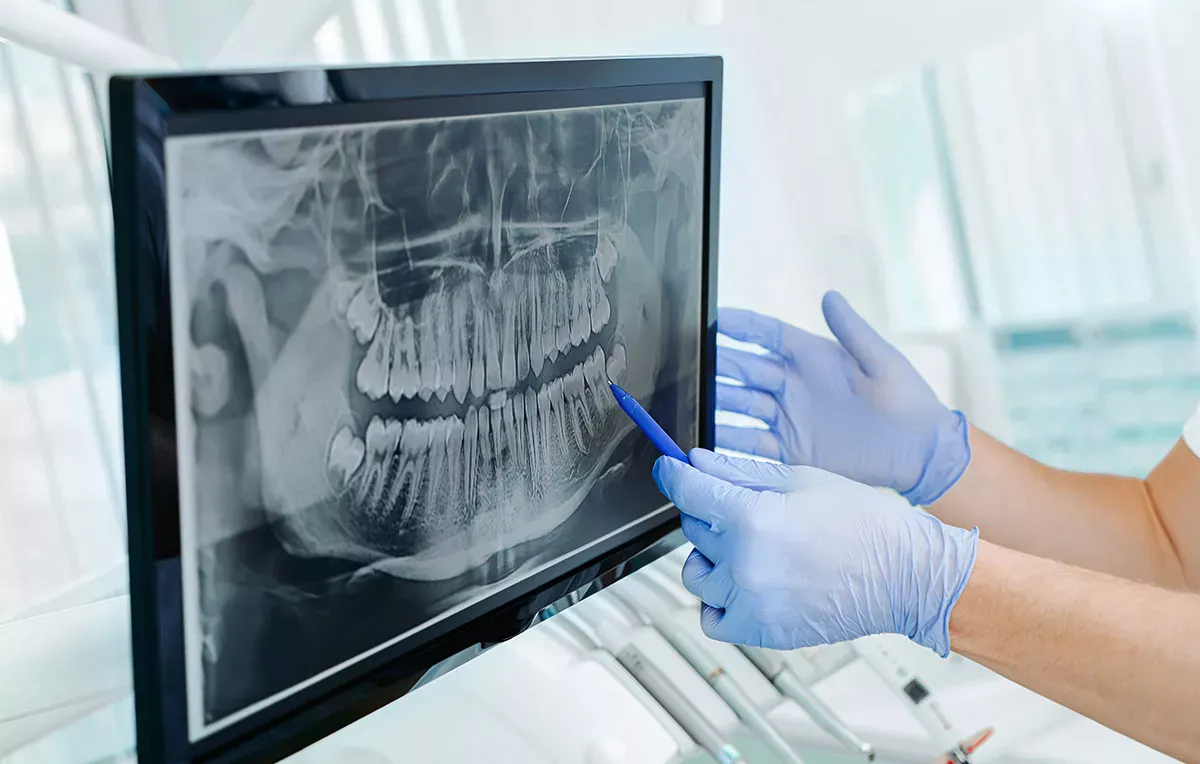
The dentist can detect inflammations, cavities, caries and other changes to the tooth or jaw substance on the X-ray because these areas are darker in colour than healthy teeth.
Are X-rays at the dentist dangerous?
When X-rays penetrate the body, they also transfer energy to the tissue, which can lead to damage either directly or through subsequent processes. However, as the human body has strong protective mechanisms, this damage can usually be repaired quickly. It only becomes problematic if the radiation exposure is too high and the self-repair mechanism can no longer cope or if unrepaired changes degenerate into cancer.
Current research indicates that the probability of this type of damage increases proportionally with the dosage of radiation. The higher the radiation exposure, the higher the risk of potentially harmful changes. In a 2012 US study, for example, scientists found that frequent X-ray examinations with a radiation dose that is no longer used today can trigger benign brain tumours (meningiomas). However, the results of this study are no reason to panic: Normal X-rays in the dental practice use a much lower dose than the cases studied, so the risk of damage is minimal these days. As an additional safety precaution, patients wear a lead apron during radiotherapy to protect their organs.
Good to know:
Restrictions apply to many examination procedures for people with pacemakers, but not for dental X-rays.
Classification of radiation exposure
However, radioactive radiation is not only produced by X-rays. We are also constantly exposed to radioactive radiation in our everyday lives. Here are a few comparisons from the patient information of the German Society for Dental, Oral and Maxillofacial Medicine to help you categorise the radiation exposure caused by dental X-rays:
A small X-ray of the teeth has an exposure of around 5μSv. In Germany, the natural radiation exposure from space and from the ground is around 2.1mSv. The annual exposure in everyday life is therefore around 400 times higher than the exposure from an X-ray. A digital 3D volume tomography, in which the X-ray tube rotates around the patient's entire head, results in an exposure of around 100μSv. This is roughly equivalent to the radiation exposure you are exposed to on a flight from Germany to Brazil and back, and is less than one twentieth of the natural annual exposure.
How often can I have my teeth x-rayed at the dentist?
Despite the low radiation exposure, dentists are required to reduce the residual risk as far as possible and avoid unnecessary x-rays. In concrete terms, this means that the dentist must decide for each individual case whether the benefit of the X-ray is greater than the potential harm.
However, we cannot give any generalised information on frequency, as different patient groups require different treatments. For example, those who have dentures in their mouth may need to have an annual update of the X-ray image, while for patients without dentures, an X-ray every two years or less may be sufficient.
Are x-rays at the dentist dangerous during pregnancy?
The risk of radiation reaching the uterus during dental x-rays is very low - especially if the patient is wearing a lead apron in the pelvic area. However, as there is a minimal residual risk, dentists generally postpone X-rays until after the birth. The growing embryo is particularly sensitive in early pregnancy, i.e. in the first few weeks. If you want to have children, it makes sense to go to the dentist before you start planning your family and to have any necessary x-rays before implantation. In urgent cases, however, it is also possible to have dental x-rays taken during pregnancy .
X-rays at the dentist are safe during breastfeeding and you do not have to take a break from breastfeeding.
Compulsory further training
To ensure that dentists are familiar with the current safety regulations for X-rays, they must attend an X-ray course at least every five years. The regulations for quality assurance in X-rays are particularly strict in Germany, Austria and Switzerland.
What types of x-rays are available at the dentist?
Not all X-rays are the same. There are various procedures for imaging teeth or the jaw area. Here is an overview:
Digital X-ray vs. analogue X-ray
The difference between analogue and digital in X-rays is similar to that in photography: just like an analogue camera, an analogue X-ray machine needs film to image the rays. In digital X-rays, imaging plates or sensors are used instead. This has the advantage that the digital images have a higher image quality and can also be post-processed using an image processing programme. For example, the dentist can adjust the brightness, increase contrasts or enlarge conspicuous areas so that they can be better assessed. The images are saved digitally so that the dentist can view them directly on the monitor. Digital X-ray images are also easier to store and pass on.
Another advantage is that digital X-rays cause significantly less radioactive radiation than analogue X-rays because sensors and imaging plates are much more sensitive than X-ray films and therefore require less radiation. Digital X-rays produce up to 90 per cent less radiation exposure. Some dental practices therefore perform dental x-rays without a lead apron . However, we would like to point out that radiation protection bodies are still in favour of patients wearing a lead apron for protection.
Most dental practices in German-speaking countries have now switched to digital X-rays.
X-ray techniques at a glance
With analogue and digital X-rays, either individual teeth or the entire set of teeth can be photographed.
Intraoral X-ray
Intraoral X-ray means that the patient has the imaging plate or X-ray film in their mouth and holds it in place by lightly biting down. This technique is mainly used when individual teeth need to be examined specifically, for example because there is a suspicion of inflammation of the tooth root or caries in the spaces between the teeth.
Good to know:
You can find out exactly what tooth decay is and how to treat affected teeth in our article:
Panoramic X-ray
Panoramic x-rays (technical term: orthopantomography) are used to obtain an overview of the entire oral cavity in order to detect abnormalities such as root inflammation, tumours and abscesses or to view the position of wisdom teeth that have not yet erupted. In contrast to intraoral X-rays, the film or sensor is located outside the mouth area. A special X-ray machine travels completely around the patient's head.
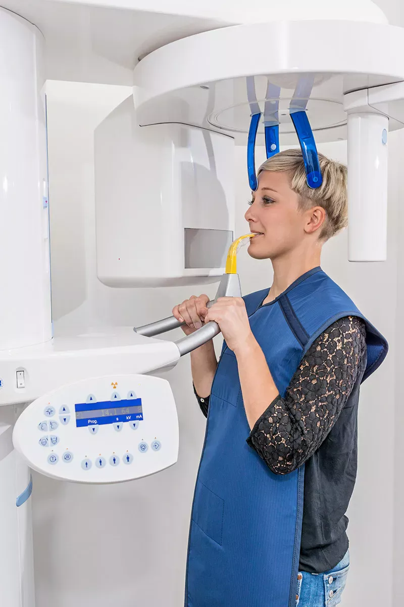
Digital volume tomography (3D X-ray)
Digital volume tomography (DVT) offers an even better overview, as it visualises the entire oral and maxillofacial region in three dimensions. This is why it is also known colloquially as a "3D X-ray". It works as follows: The device moves around the patient's head, generating around 200 to 600 fluoroscopic images, which are combined into a volume in a computing process. This produces a detailed 3D image. The advantages are that dentists can determine the bone volume and bone quality and use this information to plan implants. However, 3D X-rays are not necessary for the diagnosis of diseases.
Good to know:
What exactly are implants and what alternatives are there? Find out everything you need to know about dentures in our article:
FAQ: Frequently asked questions about x-rays at the dentist
Will I have to pay for a dental X-ray?
Both statutory and private health insurance companies generally cover the costs of analogue and digital X-rays. One exception is digital 3D volume tomography, which is not covered by statutory health insurance in Germany and Switzerland . It usually has to be paid for by patients with statutory health insurance. The costs for this are between 150 and 250 euros.
Can I request an X-ray from the dentist?
If you have changed dentist and your old dentist still took an X-ray, you have the right to request the X-ray to be given to your new dentist. Your health insurance company may also request the x-ray as proof of planned treatment. Your new dentist can also request the X-ray directly from the previous dentist. According to data protection law, your consent is not required for this.
How long must X-ray images be stored?
The retention period for x-rays of adults is ten years for dentists. X-rays of minors must be kept until they reach the age of 28.
What hygiene measures are there for x-rays at the dentist?
The parts of the X-ray machine that the patient has touched must be disinfected after each use. This applies in particular to X-ray films and sensors that patients put in their mouths.
Can I refuse x-rays at the dentist?
If the only option for you is a visit to the dentist without an X-ray, you also have the right to critically question or even refuse the X-ray. However, you should be aware of the consequences: Without an X-ray, the dentist may not be able to make the correct diagnosis in the event of complaints and may not be able to recognise abnormalities at an early stage.
Sources
Academy of Breastfeeding Medicine: ABM Clinical Protocol No. 31: Radiology and Nuclear medicine: studies on breastfeeding women.
Ärzteblatt: X-ray at the dentist as a meningioma risk.
Bayerische LandesZahnärzte Kammer: Disclosure of X-ray images.
German Dental Association: Implementation recommendations for dental radiology.
Claus, Elizabeth et al: Dental X-Rays and Risk of Meningioma, in: Cancer. 2012.
Dentlounge: Digital volume tomography (DVT).
DentNet: Digital X-ray - low radiation, many advantages.
German Society for Dental, Oral and Maxillofacial Medicine (DGZMK): X-rays at the dentist.
Hajtó, Jan : X-rays at the dentist - the facts, on: hajdo.de.
Interessenverband Zahnärztlicher Verbände in Deutschland IGZ e.V.: Das zahnärztliche Röntgen.
State Institute for Occupational Safety and Health North Rhine-Westphalia:Is it correct that a lead apron is not required to protect patients in dentistry when taking bitewing radiographs using digital X-ray technology?
Landeszahnärztekammer Baden-Württemberg: FAQ General Law.
Malitte, Hans-Joachim et al.: Strahlenschutz in der ZfP für Deutschland, Österreich und die Schweiz -Gemeinsamkeiten und Unterschiede, on: ndt.net.
Medtronic: Living with a pacemaker.
Meindentist: DVT at the dentist.
Nicol, Philipp: X-ray, on: netdoktor.de.
Petrali, Luka: Is X-ray harmful?, on: zahnarztcentrum.ch.
Rahimian, Neda: X-rays at the dentist, on: fitdentist.de.
Rheia, Nicola: Hygiene management in the dental practice, on: zmk-aktuell.de.
Rottke, Dennis: Digital volume tomography: What you need to know, on: zmk-aktuell.de.
Süddeutsche Zeitung: X-rays in the mouth: No more than absolutely necessary.
Swissmom: Visit to the dentist if you want to have children.
Dentists at Löwenplatz: 3D X-ray (DVT) in Zurich.
Future tooth: Digital X-rays at the dentist.
All websites last accessed on 27/10/2023.