Swiss premium oral care

Definition: Is there such a thing as gum cancer?
Yes, gum cancer does exist. Gum cancer is a malignant tumour that forms on the gums of the lower and upper jaw. This type of cancer is relatively rare, representing less than ten percent of all malignant tumours in the oral cavity. The technical term for gum cancer is gingival squamous cell carcinoma. Unlike other oral cavity cancers, gum cancer can be mistaken for various gum or dental conditions. Gum cancer occurs more frequently in the lower jaw than in the upper jaw.
In the majority of cases, gum cancer is a squamous cell carcinoma. This means that the tumour is located on the uppermost layer of the oral mucosa. Other types of cancer are much rarer – for example osteosarcomas, which develop in the jawbone and infiltrate the gums, or mucosal melanomas, which are often dark in colour. Early diagnosis is crucial for squamous cell carcinomas, as the tumour has a tendency to invade adjacent tissues and develop metastases in the lymph nodes. Tumour cells can spread throughout the body through the lymphatic system, thereby impacting vital organs.
In contrast to other forms of oral cancer, the gender ratio among individuals affected by gum cancer in Germany is almost equal. This type of cancer occurs only slightly more often in men than it does in women. With other types of oral cavity cancer, the male proportion can reach up to 75 percent. In men, gum cancer primarily occurs between the ages of 55 and 79, while women are more likely to develop the disease in old age, starting from the age of 75.
Symptoms: How do you recognise gum cancer?
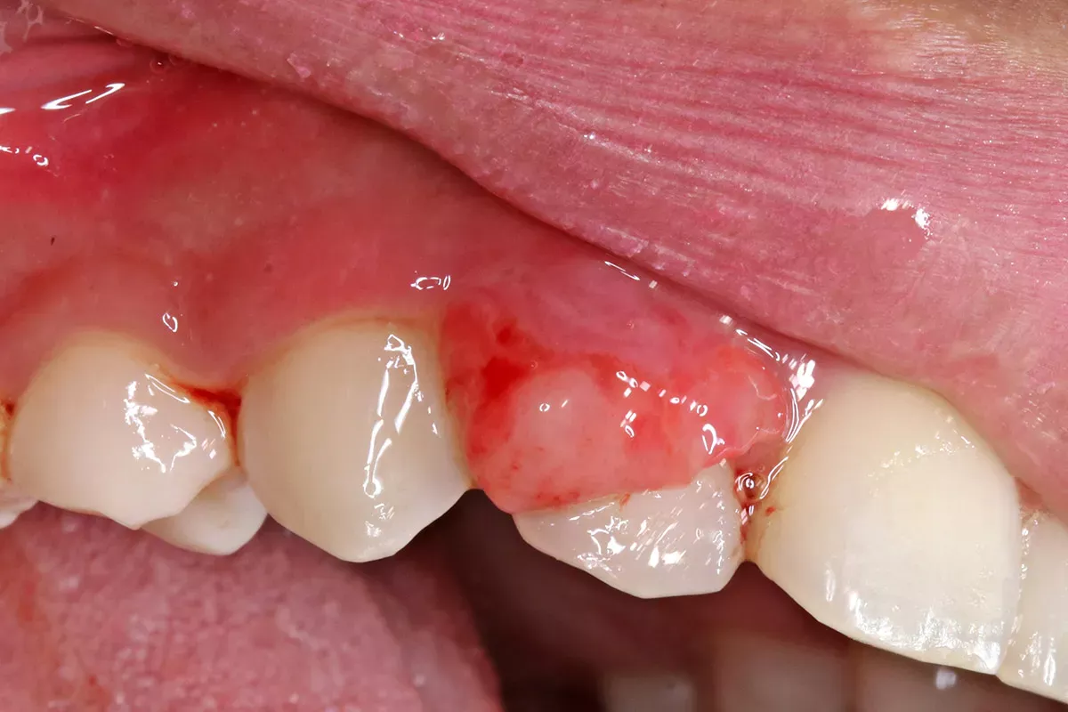
In its early stages, gum cancer often progresses without any symptoms or discomfort. However, there are some mucosal changes that – if left untreated – may develop into gum cancer. The images below depict possible preliminary stages as well as advanced gum cancer.

Possible preliminary stages of gum cancer
Leukoplakia and erythroplakia, found in various areas of the mouth, have the potential to progress into malignant tumours.
Leukoplakia
Leukoplakia is known as a condition that causes white gums. It can appear as white patches that cannot be wiped off and may cover certain areas or the entire oral mucosa. Depending on the specific form, three to 38 percent of leukoplakia cases develop into a squamous cell carcinoma if left untreated. Leukoplakia occurs when the oral mucosa exhibits a high degree of keratinisation. The primary risk factors for leukoplakia include tobacco consumption (including snus), alcohol use and poor oral hygiene.
Erythroplakia
Erythroplakia presents as velvety, bright dark red patches on the oral mucosa, which can also occur on the gums, and are much rarer than leukoplakia. If left untreated, 90 percent of erythroplakia cases develop into cancer.
Signs of gum cancer
With gum cancer, the tumour either grows out of the gum as an ulcer and initially looks like a swelling, or it grows inwards, creating a "crater". This is also referred to as an ulcerative lesion. Gum cancer is typically identified by the presence of a tumour ulcer.
Furthermore, the following symptoms may also be present:
- Bleeding gums
- Cracks in the gums
- Pain in the gums
- Loose teeth
- Sores that do not heal
- Numbness
- Dentures that no longer fit
- Ear pain
- Neck pain
- Swollen lymph nodes
In the advanced stage, symptoms of generally feeling unwell, e.g. tiredness, lethargy, loss of appetite and weight loss, also occur.
What does gum cancer look like?
In its early stages, gum cancer often appears inconspicuous, leading to potential confusion with various other dental and gum diseases.
Gum cancer can mimic the following oral and dental problems:
- Gum disease (gingivitis)
- Periodontitis
- Fungal infection
- Traumatic ulcers
- Abscesses and cysts in the gums
- Aphthae
An incorrect diagnosis and premature treatment by a dentist can have detrimental consequences. Dentists should, therefore, perform a biopsy on suspicious areas in the mouth to rule out cancer.
Causes
Gum cancer develops when cells with altered genetic information are not identified and destroyed by the immune system and start to multiply. Since tumour cells divide faster than normal ones, they gradually displace more and more healthy cells, which allows for tumour ulcer progression. Unfortunately, it is not yet known exactly how these cells mutate. There are, however, a number of risk factors that favour the development of gum cancer.
Risk factors
Smoking tobacco and drinking alcohol on a regular basis are the main risk factors for oral cavity cancer. Although gum cancer is a form of oral cavity cancer, the link here is comparatively weaker than with other types of cancers. While it is true that gum cancer was more prevalent during periods when the use of chewing tobacco and tobacco consumption was widespread, it is important to note that individuals who have never used tobacco can still develop gum cancer.
Numerous studies in Sweden, the primary exporter of snus, a smokeless tobacco, have thus far found either no link or only a very slight association between the use of snus and oral cavity cancer. Other studies, however, have confirmed a direct link between snus consumption and the development of leukoplakia, a potential preliminary stage of gum cancer. Mucosal thickening, which appears white, often occurs at the application site and can develop into a tumour.
Poor oral hygiene and ill-fitting dentures, which cause chronic inflammation, are also considered possible risk factors.
Good to know:
Everyone can prevent poor oral hygiene. Find out how to brush your teeth correctly and which routine you should follow here:
Diagnosis: Who diagnoses gum cancer?
It is common for individuals to seek advice from a dentist when they notice something unusual about their gums or oral health. However, gum cancer is also frequently discovered incidentally during routine check-ups or professional dental cleaning sessions by dentists or dental care professionals.
If other gum diseases have been ruled out or the diagnosis is uncertain, the dentist may refer the patient to a specialist, such as an ear, nose and throat (ENT) specialist or an oncologist (cancer specialist). The first step in diagnosing cancer is a biopsy during which the doctor takes a tissue sample from the affected area, which is then tested in a laboratory to determine the presence of tumour cells. If the suspicion of cancer is confirmed, various imaging techniques are employed:
- Computed tomography (CT) scan
- Magnetic resonance imaging (MRI) scan
- Ultrasonic scan of the lymph nodes
- X-ray examination of the upper and lower jaw
A doctor determines which examinations are necessary. The specialist then uses the examination results to classify the tumour. Several factors influence this classification, including the size of the tumour, its invasion into surrounding tissues, the presence of metastases in lymph nodes or other organs and its rate of growth.
Good to know:
Find out exactly how tumours in the oral cavity are classified in our main article:
What does gum cancer treatment entail?
Gum cancer typically requires surgical removal of the tumour. Depending on the stage, size and location of the tumour, radiotherapy or chemotherapy may be administered before or after surgery to prevent tumour recurrence. For example, radiotherapy alone might be sufficient to cure small tumours.

The most effective treatment plan is determined by a team of specialists from various medical disciplines who collaborate in meetings known as tumour conferences to discuss each individual case.
The treatment of gum cancer has three specific goals:
- Remove the tumour completely
- Maintain the function and appearance of the mouth as best as possible
- Prevent recurrence of the tumour
Good to know:
Cancer treatment unfortunately has negative impacts on both dental and oral health. Read about the exact side effects and how you can minimise them in our article:
Surgery
The surgical procedure to remove the tumour can vary significantly based on its size: Small, localised tumours can be surgically excised relatively easily. However, in cases where a significant amount of gum and bone needs to be removed, resulting in inadequate tooth support, tooth extraction may be necessary.
When the tumour has infiltrated the jawbone, a complex surgical procedure is necessary, often involving the removal of a part of the jaw. In one or multiple reconstruction procedures, the surgeon grafts bone and tissue from the patient's own body, often extracting parts of the fibula or shoulder bone, to rebuild the jaw. Implants or prostheses are used to fully restore chewing and speech capabilities.
If the tumour has already metastasised to the lymph nodes or is suspected of having done so, they also have to be excised. This type of surgery is called a neck dissection. Explanation: Neck dissection refers to a surgical procedure in which either all the lymph nodes of the neck or the lymph nodes on one side of the neck are removed as part of tumour surgery in the head and neck region.
Good to know:
More information on the various treatment methods can be found in our main article on oral cavity cancer:
Prognosis: Can gum cancer be cured?
Like most types of cancer, gum cancer is curable if it is recognised early enough. The chances of survival and recovery depend greatly on the stage of the tumour and whether metastases have already formed. Other factors that play a role include the age of the patient at the time of diagnosis, the size of the tumour, the involvement of the lymph nodes and the chosen treatment method.
In general, oral cavity cancer is curable in 80 to 90 percent of cases if detected at an early stage. However, only 30 percent of all oral cavity cancers are detected early on. To prevent a tumour from developing unnoticed in your mouth, you should therefore go for regular dental check-ups.
The prognosis worsens as the tumour advances. Unfortunately, we are unable to make any general predictions regarding the duration and effectiveness of treatments, as this varies from case to case.
Good to know:
Self-examine your mouth regularly for any suspicious signs or changes. Discover how to do this exactly in our main article on oral cancer:
Preventative measures: What you can do to prevent oral cavity cancer
Sources
Beisel, Sebastian: Den Tumor erkennen, bevor er entsteht!, at: zwp-online.info.
Demarco, Cynthia: Cancer of the gums: 9 things to know, at: mdanderson.org.
Deutsche Krebshilfe: Krebs im Mund-Kiefer-Gesichtsbereich (Die blauen Ratgeber).
Deutsche Krebsgesellschaft: Klassifikation von Tumoren (TNM-System & Grading).
Driemel, Oliver et al.: Erkennung oraler Risikoläsionen in der zahnärztlichen Praxis. Zahnarztexemplar.
Gesellschaft für Zahngesundheit, Funktion und Ästhetik (GZFA): Formen der Parodontitis.
Gupta, Renu et al.: Gingival squamous cell carcinoma presenting as periodontal lesion in the mandibular posterior region, in: BMJ Case Reports. 2014.
Leitlinienprogramm Onkologie (Arbeitsgemeinschaft der Wissenschaftlichen Medizinischen Fachgesellschaften e. V. (AWMF), der Deutschen Krebsgesellschaft e. V. (DKG) und der Stiftung Deutsche Krebshilfe: Patientenleitlinie Mundhöhlenkrebs.
Lee, Jang-Jaer et al.: Gingival squamous cell carcinoma mimicking a dentoalveolar abscess: report of a case, in: Journal of Endodontics. 2007.
Leitlinienprogramm Onkologie: S3-Leitlinie Diagnostik und Therapie des Mundhöhlenkarzinoms.
Memorial Sloan Kettering Cancer Center: Gum Cancer.
Molina, Ana Paula et al.: Gingival squamous cell carcinoma mimicking periodontal disease, in: International Journal of Periodontics & Restorative Dentology. 2011.
Ramesh, Roshi et al.: Oral squamous cell carcinoma masquerading as gingival overgrowth, in: European Journal of Dentistry. 2017.
Robert Koch Institut: Datenbankabfrage, at: krebsdaten.de
Sieber, Andreas et al.: Snus und die Beeinträchtigung der Mundgesundheit, in: Swiss Dental Journal. 2016.
Uniklinik RWTH Aachen: Mundschleimhautveränderungen.
Universitätsklinikum Düsseldorf: Mundschleimhauterkrankungen.
All websites last accessed on 6 September 2023